Who first thought of a gait graph?

Quite out of the blue Jenny Kent from Headley Court asks if I know where the gait graphs we know today come from. She was particularly interested in where the idea of time normalising data to the gait cycle originated. I have to admit I just don’t know.

Braune and Fischer, working at the end of the 19th century, certainly plotted a number of gait variables against time, most for swing but a few for more than a gait cycle. All the graphs I can see though plot these against time rather than a percentage of the gait cycle and the data for more than a gait cycle doesn’t appear to be plotted in relation to the gait events at all.

The first group that I can find that present variables on graphs with the time axis labelled as % gait cycle is Inman’s group working in Berkeley in the early 1950s.

Inman time normalisation

Data scanned a long time ago from one of the outputs of the Berkeley group – not sure which.

Can anyone provide any earlier examples?

.

This made me think about other features of our standard gait graphs. Who first proposed plotting data from a patient against normative reference data depicted as a mean and range based on the standard deviation?

I remember that when the Vicon Clinical Manager software came out in 1992 that it assumed that all data was normalised to the gait cycle (the data was actually stored in a .gcd file on this assumption). The software only allowed three traces to be plotted on any graph so the common practice was to plot the mean of the reference data along as one right and one left side trace for each patient. I think the practice of plotting several (three!) traces from each side separately to assess measurement variability probably dated to this time as well. I don’t remember the standard deviations being plotted but this may just be my memory (the standard deviation values could certainly be stored in the .gcd file).

I also remember being impressed by teaching material from Newington and Gillette Hospitals (Gage, Davis and Ounpuu) which plotted the standard deviation ranges from quite an early stage. Looking up some of their early papers I find that  Sylvia’s 1995 paper contains sample patient data plotted against the standard deviation ranges. (Unfortunately the quality of this figure in the .pdf file I have is too poor to be worth reproducing here).

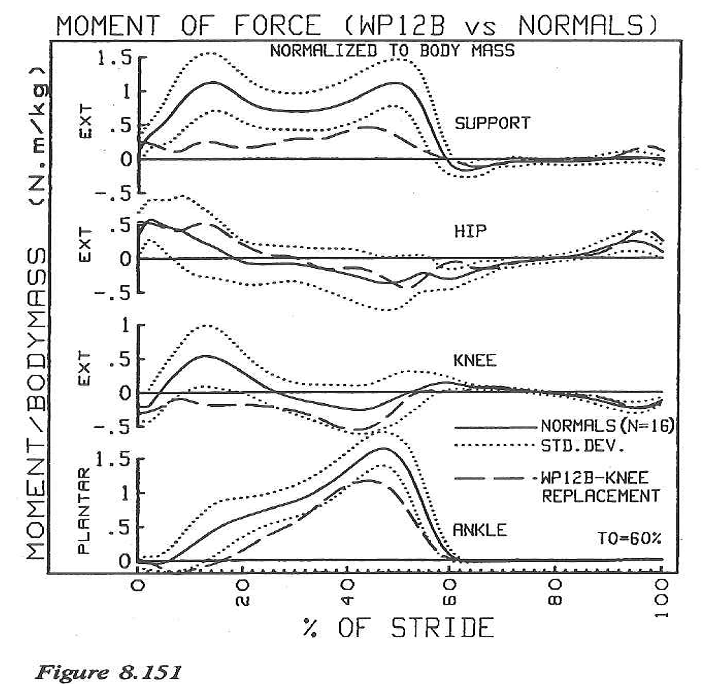
Sylvia moved to Newington from Waterloo so I wondered how David Winter had plotted his data. Sure enough in the final chapter of The Biomechanics and Motor Control of Human Walking (1991) entitled “Assessment of pathological gait” are a series of graphs showing gait variables from a patient with a knee replacement plotted against the mean and standard deviation from a reference population. (This book was an adaptation of an earlier one form 1987 which I don’t have access to and I’d be interested to know if these graphs were included in that as well).

 winter gait graphs

I’d like to suggest that this might be the earliest example of gait graph as we use them today – or has anyone got any earlier examples?

Of course tracing ideas back like this is a slightly ridiculous activity because such graphs  often appear in publications only after having been used more generally for a considerable period. Just because they first appear in print from one team does not necessarily mean that they originated there!

.

Braune, W., & Fischer, O. (1987). The Human Gait (P. Maquet & R. Furlong, Trans.). Berlin ; New York: Springer-Verlag.

Klopsteg, P. E., & Wilson, P. D. (1954). Human Limbs and their Substitutes. New York: McGraw-Hill.

Ounpuu, O., Davis, R., & Deluca, P. (1996). Joint kinetics: Methods, interpretation and treatment decision-making in children with cerebral palsy and myelomeningocele. Gait and Posture, 4, 62-78.

Winter, D. (1991). The biomechanics and motor control of human gait: Normal, Elderly and Pathological (2nd ed.). Waterloo:: Waterloo Biomechanics.

Why bother with gait classification?

There is a sector of our community that sees classification as the holy grail of gait analysis. If only we could divide our patients into neat little categories then our problems will be solved. But why do we want to classify patients?

The most obvious reason for categorising patients into groups would be if genuinely different groups existed. But do they? In an earlier blog I’ve written about the GMFCS and pointed out that there is absolutely no reason to believe that children with cerebral palsy fall into distinct categories of severity – they almost certainly lie on a continuous spectrum.  I suspect that the same is true of gait patterns. Indeed in work that Fiona Dobson completed for her PhD thesis but never published we used a technique called multi-dimensional scaling to look for evidence of clustering of gait patterns in children with hemiplegic cerebral palsy. We couldn’t find any. Whichever way we looked at the data it looked like a random scatterplot.

So if there is so little evidence that gait patterns fall into distinct categories why are we so obsessed by it?

I suspect that classification systems are assumed to help clinicians – particularly less specialist ones than are employed in our gait analysis services. If we can simplify understanding of gait to a small number of discrete categories then we’ll be able to teach people to recognise these. If we can describe management plans for the different categories then maybe we can even teach people to treat the patients.

There are problems though.  The first one is that the classification systems don’t seem to work – clinicians can’t agree on what category a gait pattern should be placed in. Fiona Dobson, in one of the few studies to assess reliability of a classification in a centre outside that in which it was developed (Dobson et al,2007), looked at the agreement of 16 clinicians in rating gait patterns of 34 children with hemiplegic cerebral palsy . On the basis of video data, only one pattern was put in the same category by all clinicians and in over a third there was less than 50% agreement. An important feature of this study was that the 16 clinicians were all specialists in gait analysis of children with cerebral palsy. What would the results have been like if the non-specialist clinicians, who presumably have most to gain from classification systems, had been recruited to the study? Results were a little better when gait analysis data was used as a basis for classification, but then the clinicians for whom classification is likely to be most useful don’t generally have access to gait analysis.

Another issue is that gait patterns are almost certainly a combination of characteristics of the child and of how they have been managed previously. The Winters, Gage and Hicks (1987) classification of hemiplegia intentionally excluded children who had had previous surgery but the classification of diplegia by Rodda et al. (2004) included children with prior calf surgery (because there were so many of them). We never quantified it but it appeared to us that fewer and fewer children in Melbourne fell into the Rodda “crouch” category over time which we attributed to more conservative early surgical management and the wider use of Botulinum toxin. If this is the case then classifications schemes will have to be sufficiently generalizable to cover the effects of different management practices or maybe we need a number of different classification schemes depending on different management practices in different parts of the world.

Developing and validating classification schemes is a considerable undertaking. Earlier schemes were generally based on the enlightened but essentially subjective opinions of leading clinicians. In an era of evidence-based practice this is no longer satisfactory and widespread consultative processes (such as a Delphi process) would now be needed to convince the clinical community of any new scheme.  Similarly were existing schemes have been validated this has tended to be on the basis of repeatability studies based within the teams who developed the scales and typically using gait analysis techniques that may not be available to those who are most likely to benefit from the classification. Convincing validation of new schemes will need to be more comprehensive. Such an investment of time and effort would be perfectly justified if we are certain that the schemes will be useful – but we do need to be certain.

Validation is made considerably more complex by the fact that the classification systems do not fit the underlying reality. How can you interpret reliability studies designed to test the competence of clinicians to ascribe gait patterns to categories when there is genuine ambiguity in whether any individual child actually fits a particular category? A similar issue arises with the usefulness of the scheme for less experienced clinicians. Many children will either be on the borderline of categories or may not fit cleanly into categories at all. Explaining how to deal with such exceptions to generalist staff may lead to more confusion not less.

So is there an alternative? Well I’d like to float the potential of what I call profiling. Rather than select a number of discrete groups into which gait patterns can be assigned we define aspects of the gait pattern that vary across the patient population. We can then score or grade the different aspects separately. This allows us to accept that there is a continuous distribution of gait patterns and that also that gait might vary independently across the different aspects rather than suggesting that there is necessarily grouping of the aspects as is required by classification schemes.

We presented a paper proposing this for the gait of children with hemiplegic cerebral palsy at the Sydney CP meeting in 2008 (Tirosh, Dobson et al.) but never took the ideas any further. A factor analysis based on movement analysis profile scores suggested that 85% of the variability could be explained by 5 factors which reflected:

  • distal sagittal plane features (knee flexion and ankle plantarflexion combined),
  • leg length discrepancy (anatomical and functional),
  • hip rotation,
  • hip flexion
  • foot alignment.

These factors all make clinical sense as does the idea that hemiplegic gait should be categorised by assessing how far each of these different aspects contributes to the gait pattern of an individual child rather than trying to assign the gait pattern to one of a small number of categories. Perhaps more importantly it appears to me (and I admit this is a subjective opinion) that the underlying model of gait patterns varying along a continuum in a number of different dimensions matches reality more closer than a model assuming that gait patterns fall into distinct and recognisable categories.

.

Dobson, F., Morris, M. E., Baker, R., & Graham, H. K. (2007). Gait classification in children with cerebral palsy: a systematic review. Gait and Posture, 25(1), 140-152.

Rodda, J. M., Graham, H. K., Carson, L., Galea, M. P., & Wolfe, R. (2004). Sagittal gait patterns in spastic diplegia. Journal of Bone and Joint Surgery. British Volume, 86(2), 251-258.

Winters, T. F., Jr., Gage, J. R., & Hicks, R. (1987). Gait patterns in spastic hemiplegia in children and young adults. Journal of bone and Joint Surgery – American, 69(3), 437-441.

What the ICF?

If readers don’t recognise this diagram then they should. It comes from the World Health Organisation’s International Classification of Functioning , Disability and Health (ICF). It proposes a model within which to consider how any health condition affects a person’s life. It’s becoming an important tool to plan research on the assumption that interventions to improve people’s walking ability should be assessed over a range of levels.

Image

It assumes that the affect that a health condition has on a person’s life depends on a balance between attributes of the health condition itself and a range of contextual factors which can relate to the environment in which the person lives of be personal characteristics. Thus the life of a person who cannot walk (health condition) will be affected by environmental factors such as whether they have a wheelchair and whether the places they want to visit are wheelchair accessible and personal factors such as motivation or coping abilities.

Three levels are used to describe the person relate to the body or a body part (body structure and functions), the whole person (activity) and the whole person within a societal context (participation). Factors affecting body structure and function interact with those affecting  activity (this is a two way interaction – horizontal arrows). Those affecting activity also interact with those activity interact with participation. Note that there is no direct interaction between body structure and function and participation – I’ll pick this up in a later post).

Another aspect of the classification is a distinction between capacity, what a person is able to do within a standardised environment, and performance, what they actually do in their current environment. Thus we have come across children with disability who have the capacity to walk with crutches, but perform as wheelchair users because some schools consider this to be safer.

If we just look at this diagram it’s not at all clear where walking fits in. Most people don’t look any further than the diagram which means there is some ambiguity in the literature considering walking within the context of the ICF. The ICF is not limited to the diagram however and contains an entire classification system (as the name suggests!). Perhaps the easiest way to get your head around the classification itself is to use the illustrated version.

icf pics

Images from the illustrated ICF (click on picture to take you there)

In this the gait pattern is listed as item b770 in the Neuromusculoskeletal and Movement Related Functions chapter. It thus becomes very clear that walking as measured by a gait analysis service is an example of a body function (and more specifically that the measures are of capacity – you don’t get a much more standardized environment than a gait analysis laboratory).

Walking as it occurs as part of everyday life is defined as an activity within the Mobility chapter of the ICF. It is further divided into walking over different distances (d4500 & d4501), on different surfaces (d4502) and around obstacles (d4503). For most people walking will be the main method of moving around the home (d4600), in other buildings (d4601) and for short distances outside the home (d4602). By definition this activity cannot be measured within the gait laboratory but questionnaires or data logging devices can be used to obtain the information. The Functional Mobility Scale (Graham, 2004) , for example, assesses dependence on walking aids over three distances, 5m, 50m and 500m which fits nicely onto categories d4600, d4601 and d4602 (although I don’t think any of the team were conscious of this when the scale was being developed).

How a person’s participation in society will clearly be affected by their walking ability but is not covered by any specific codes within the ICF.

.

PS

The history of the development of the ICF (and its predecessor, the ICIDH) makes interesting reading. It is the result of an ongoing discussion as to whether health should be primarily viewed using a medical or social model (see the  Beginner’s Guide or the article in the International Encyclopaedia of Rehabilitation). The two classifications also marked a transition from the WHO’s original focus on preventable deaths (which are still the primary health concern in many of the world’s poorest countries) to what represents health (which is much more comfortable for those of us in richer countries). This rarely gets written about but then again most people doing the writing are doing so from within the richer countries!

.

PPS in writing this post I spotted the first error in my book – the horizontal arrows are missing from the version of the ICF diagram (Figure 9.1). Do let me know if you find others.

.

Graham, H. K., Harvey, A., Rodda, J., Nattrass, G. R., & Pirpiris, M. (2004). The Functional Mobility Scale (FMS). J Pediatr Orthop, 24(5), 514-520.

Kinetics of the inverted pendulum

One of my first posts was about the inverted pendulum and in it I promised a follow-up that I never delivered. So here it is. I commented that for all there is a lot of talk about the inverted pendulum there is little understanding of what it is and what it’s characteristics are. I’ll focus on the kinetics today.

The graph below shows the vertical component of the ground reaction under an inverted pendulum (Anderson and Pandy, 2006). You can work out the shape the curve must have from basic physics. Early on the pendulum is rising as it moves towards the vertical. As it is does so it gains potential energy and must be losing kinetic energy. Its upward velocity is thus reducing so it has a downward acceleration (i.e. an upward deceleration). If the overall force is acting downwards then the ground reaction (up) must be less than bodyweight (down). As the pendulum moves towards its highest point along a circular arc it rises less slowly, decelerates less, so the ground reaction must get closer to bodyweight.

inv pen GRF

Once it is over the highest point it starts to lose height, and accelerate downwards. Again this requires a downwards force so again the ground reaction (up) must be less than bodyweight (down). The further the mass goes around the circular arc the more quickly it loses height, the more it accelerates, so the ground reaction must be a decreasing fraction of the ground reaction. Easy eh! Appliance of science and we can predict the curve above.

The interesting thing here is that the vertical component of the ground reaction under an inverted pendulum is always less than its own weight. The inverted pendulum may be an excellent mechanism for carrying a mass from one point to another but its a pretty hopeless one for supporting that mass. On reflection this should be obvious because the vertical component of velocity is upwards at the start and downwards at the end and thus the nett acceleration during the movement is downwards and the average force must be less than bodyweight.

If the average force is less than bodyweight then you can’t possibly have a viable walking pattern simply by stringing a series of inverted pendulums together no matter how good the drawings of the kinematics look.

There are two mechanisms by which we get over this. The first is that we use our muscles so that the the ground reaction does not just mimic the mechanics of a passive inverted pendulum. In the figure below the ground reaction is under an inverted pendulum (solid line) is plotted against Winter’s data (1991)  for the vertical component of the ground reaction (grey band) from the middle of one double support phase to the next. The characteristics double bumps of the ground reaction clearly increase the average vertical force (all forces are plotted as % bodyweight).

GRF

This isn’t the whole story however. If you look more critically at this data you will see that the average vertical component of the ground reaction under the body is still considerably less than bodyweight (about 10% less) for most of us. The peaks aren’t much higher than bodyweight and they don’t last that long. How can we walk around and not support out own bodyweight?

The answer lies in two words, “double support”. During double support the forces  under both limbs combine to exceed bodyweight. The largest total vertical component of the ground reaction is actually in mid double support when relatively modest looking ground reactions under both limbs combine (you can see a graph of this in an earlier blog). By allowing the two ground reactions to combine like this we are able to rely on an inverted pendulum like movement to move the body forwards whilst achieving an average total ground reaction equal to bodyweight – a fundamental pre-requisite of cyclic walking.

A double support phase is thus an essential requirement of a gait pattern based on an inverted pendulum. It’s interesting that modelling the body as a simple inverted pendulum leads to a prediction that double support needs to last for 15% of the gait cycle. The actual value is, of course, 10%. That’s not a bad guess for such a simple model.

I put these ideas in a slightly wider context in one of the screencasts in the series “Why we walk the way we do. The whole series is linked to on my Videos page.

.

Anderson, F. C., & Pandy, M. G. (2003). Individual muscle contributions to support in normal walking. Gait Posture, 17(2), 159-169.

Buczek, F. L., Cooney, K. M., Walker, M. R., Rainbow, M. J., Concha, M. C., & Sanders, J. O. (2006). Performance of an inverted pendulum model directly applied to normal human gait. Clin Biomech (Bristol, Avon), 21(3), 288-296.

Winter, D. (1991). The biomechanics and motor control of human gait: Normal, Elderly and Pathological (2nd ed.). Waterloo:: Waterloo Biomechanics.

The importance of objective outcome measurements

This post was stimulated by a presentation given to the GCMAS by Nancy Lennon of the A.I. Du Pont Hospital in Delaware. She presented data on measured activity levels over the year following major orthopaedic surgery for children with cerebral palsy. Her data came from a case study and showed how the patient’s activity levels fell markedly at 3 months after surgery before picking up through the rest of the post-operative year.

The graph below is an update of one I prepared for a lecture on Outcome Measures at the Melbourne Gait Courses last year. It puts together data from a range of sources to suggest a time history for the Gross Motor Function Measure (GMFM, Russell et al., 1989)  for a child with cerebral palsy who has single event multi-level surgery (SEMLS) at the age of 10. The data points are taken from Thomason et al (2013) and represent average GMFM scores for a cohort of children at baseline (blue) and 12, 24 and 60 months (green) following SEMLS.  Before surgery I’ve assumed that the data follows the latest GMFM curves (Hanna et al., 2009) to arrive at the baseline value.

GMFM

The red point is invented. It is an estimate of the GMFM a child might record if assessed on coming round after surgery in a hospital bed with below knee casts. The actual value is not particularly important but seems reasonable when I glance through the GMFM manual. I’ve then extrapolated the curve from this point through 12, 24 and 60 month data points.  Having seen the videos of kids coming back for 3, 6 and 9 month follow-up after such surgery whilst in Melbourne I don’t think the time course over the first year is too far away from reality. I’ve finished off the curve assuming that it follows the known GMFM data (Hana et al., 2009).

First thing to point out is that average GMFM score at one year is almost exactly the same as at baseline and the maximum GMFM is recorded at two years following surgery suggesting that the one year follow-up may be a little early to assess outcomes.

The point I really want to make though is that if you look at this graph the biggest feature is not the improvement from pre-op to 12 or 24 month status. It is the drop in function immediately after surgery and the improvement back to baseline at 12 months. This has the potential to impact on patient, family and clinical perceptions of outcomes. If the dominant memory of the surgery is of the condition the child was in immediately afterwards, then the perception may well be of the change following surgery as being represented by the difference between the green points and the red point which might lead to a much more positive view of outcomes than a more scientific comparison with the blue point. Particular caution may have to be exercised in interpreting the results of subjective or semi-subjective assessments such as heath related quality of life questionnaires or informal assessment of outcomes.

Final point is that there are a multitude of reasons for performing such surgery and assessing outcomes on the basis of any one measure in isolation is inappropriate. I’ve plotted this data to make a particular point about the time course of recovery not to make any general conclusions about the effectiveness of the surgery. Gait Profile Scores (Baker et al., 2009) reflecting the quality of the gait pattern improved by over 30% in the same cohort for example.

.

Russell, D. J., Rosenbaum, P. L., Cadman, D. T., Gowland, C., Hardy, S., & Jarvis, S. (1989). The Gross Motor Function Measure: a means to evaluate the effects of physical therapy. Developmental Medicine and Child Neurology, 31(3), 341-352.

Thomason, P., Selber, P., & Graham, H. K. (2013). Single Event Multilevel Surgery in children with bilateral spastic cerebral palsy: a 5 year prospective cohort study. Gait Posture, 37(1), 23-28.

Hanna, S. E., Rosenbaum, P. L., Bartlett, D. J., Palisano, R. J., Walter, S. D., Avery, L., & Russell, D. J. (2009). Stability and decline in gross motor function among children and youth with cerebral palsy aged 2 to 21 years. Dev Med Child Neurol, 51(4), 295-302.

Baker, R., McGinley, J. L., Schwartz, M. H., Beynon, S., Rozumalski, A., Graham, H. K., & Tirosh, O. (2009). The gait profile score and movement analysis profile. Gait Posture, 30(3), 265-269.