muscles

How long is a piece of string?

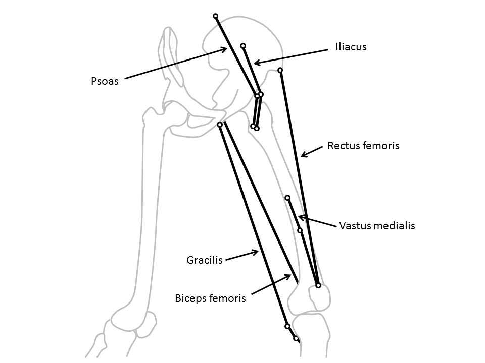
Lurking somewhere on this blog-site is software that Vicon users can download to calculate “muscle lengths”. It’s based on calculating the distance between the origin of a muscle on one segment and its insertion on another as illustrated in the diagram below (taken from my book). Some of the muscles (such as the rectus femoris) can be best represented as a straight line between the origin and insertion, whereas others (such as psoas or iliacus) have to pass around bones and may be better represented by including a “via point” along the path and adding the lengths of the two lines thus created.

muscle lengths

Clearly as the joint or joints linking the relevant segments move this distance changes and you can thus plot muscle length on gait graphs in the same way that you can plot any other gait data. The technique has been around for a very long time but has been particularly popular since Scott Delp’s work on SIMM in the late 1980s.

Recently Jussi has added a comment to the page first saying that he’s got the software working (good) but then that a “somebody” has suggested “that ‘point-to-point’ muscle length models tend to be inaccurate, and a joint angle/moment arm based methods would be more accurate”.

This is quite an interesting comment because I don’t really consider this technique as “accurate”. The technique is based on a rather crude scaling of one set of origin and insertion coordinates. We don’t really know how consistent these are across healthy individuals and certainly not  how they are affected in people with the sorts of conditions that we generally assess in clinical gait analysis (particularly those with bone, joint or muscle deformity). Further the calculations are dependent on the assumptions you make about how the joints move and ultimately on the accuracy of the joint angle measurements. All in all this is probably best described as a technique to “estimate” muscle length rather than to “calculate” it.

My general advice for clinical interpretation is that if you are dealing with single joint muscles then the muscle length graphs don’t really tell you much that you can’t already see on the joint angle graphs. Generally as a the joint extends the extensors get shorter and the flexors get longer (and vice versa) and the muscle length graph looks extremely similar to the corresponding joint angle graph (but with different units). Given that the actual calculations of muscle length are subject to so many assumptions you might as well work directly from the joint angle graphs.

The multi-joint muscles are different though because the muscle length depends on the orientation of both joints and the separate moment arms of the muscle about each. It is thus virtually impossible to assess how the muscle length varies through the gait cycle. In this case muscle length graphs are the only sensible way of getting an insight into how a muscle is behaving and can be valuable despite knowing that the actual values are only estimates. At least they are consistent estimates so that there is some sense in comparing the data you estimate for a patient against normative data which you have estimated using the same modelling procedure.

The most obvious example of this is in considering hamstrings length in children with cerebral palsy. It is extremely tempting to see a bent knee and assume that the hamstrings must be short. The “obvious” surgical response is to lengthen them. In many kids, however, the hip is also flexed and, because the moment arm at the hip is greater than that at the knee the muscles is often actually considerably longer than “normal“. This would suggest that surgical lengthening is inappropriate. Scott Delp and Alison Arnold drew the attention clinical community to this nearly twenty years ago and if there is one good reason for including muscle length estimates in gait reports for kids with CP then this is it. The data doesn’t have to be that accurate to be a reminder to surgeons that this is an important issue.

In direct response to Jussi’s question I don’t think its possible to say whether “point-to-point” or moment arm based calculations are more accurate. The calculations are affected by different factors and its not at all clear whether either is superior in general. The accuracy of whichever technique you use will be dependent on the quality of the input data (point coordinates or moment arms). As pointed out above accuracy is limited by a number of other factors and some of these may be more important than the choice of technique. Perhaps most importantly I’m not aware of any research that has ever been done to assess the accuracy of any muscle length calculations (though there is at least one that investigates the  difference in results from a using a number of techniques ).

Of course this assumes that muscle lengths are being used clinically to understand why a patient walks the way they do. Anyone wanting to use them for more technical purposes perhaps in the generation of more advanced muscuol-skeletal models really needs to develop an in depth appreciation of all these factors for themselves.

.

PS Just to avoid the terminology police its worth reminding people that what almost everyone refers to as “muscle length” is actually the musculotendinous unit length. Maybe this is something that should have been added to my rant on terminology last week.

Advertisement

Passive resistance

I’ve recently been looking at how muscles work in a little more detail than  I have for some time. In particular I’ve been looking at the so called Hill type models which place the contractile unit in series or parallel with various elastic or viscous elements. I say “so called” because when I look at Hill’s classic paper   the model is implicit rather than explicit. This probably explains why such a wide family of models are all described as Hill type models (note that a significant number of recent papers refer to the wrong classic paper which doesn’t help).

The model I’m familiar with has the contractile element in parallel with one elastic element (the parallel elastic element or PE) and both in series with the serial elastic element (SE) as summarised by the figure below.

The serial element is principally comprised of any tendon through which the muscle is attached to a bone. The parallel element is less well understood. For a long time it was assumed that this was primarily extracellular but we now know that titin also contributes within the sarcomere. Whilst in this narrative Hill was somewhat ambiguous about how the PE and SE were aligned he did publish the now famous graph of the length tension relationship for muscle (below) which clearly shows the additive nature of the contractile and parallel elastic elements which can only be a consequence of the two being in parallel.

Hill graph

Graph from Hill’s original paper

What has interested me from my recent reading has been that if you look at different sources you get quite different estimates of the amount of overlap between the passive and active components. If we assume that the location of the active curve is in the right place then the green curve is displayed shifted either to the left or right. The graphs below are all taken from different web-sites. The overlap between active and passive curves gets less as you go from left to right. So which is correct?

Hill graphs


I’ve reduced the size of these but if you were to look at the bigger versions you’d see that most look as if they have been hand sketched and don’t give a great deal of confidence that they are based on any real data. It’s not actually clear from Hill’s paper whether his passive curve is to illustrate the concept or is a represents real data (these data aren’t presented anywhere else within the paper which is principally on the mechanics of active contraction). Lieber (p51), however, presents data  (from Wang et al. 1993) suggesting that Hill’s passive curve may not be too far out. (Of course it may be, particularly if part of the passive curve is attributable to extracellular serial components, that the overlap varies from muscle to muscle or across species).

But does it matter for healthy walking? When anyone performs a physical examination of a healthy person they can generally move the joints through a significantly greater range of motion than is required for normal walking with minimal resistance. When I say minimal resistance we need to remember that the forces exerted by the major muscles during walking can be several multiples of bodyweight  (Hoy et al., 1990). This is strong evidence that the passive elastic element never comes into play during walking. We could probably get away with a simpler model that doesn’t include it at all (that just has a muscle in series with a tendon).

If we look at Hill’s graph again this makes sense as the passive load doesn’t exceed the maximum active load until the fibre is about a third longer than its resting length and there won’t be very many muscles in which the muscle extends by more than this during walking (although care is needed in pennate muscles where the change in muscle belly length may underestimate the change in fibre length). This is largely confirmed by recent data from the Stanford group (Arnold et al. 2013) which  estimates that the rectus femoris is the only muscle in which fibre lengths exceed this length during walking.

Of course this is only true for healthy walking. One of the reasons for conducting a physical examination of our patients is because we know that many of them do have passive contractures of the muscles which limit joint movement significantly. This suggests that the overlap between the active and passive curve in their muscles may be significantly different to that in the rest of us. I’m not aware of any experimental work however to have tested this (anyone know of any?).

What are the hip adductors for?

Various things have made me think about muscles this week and an issue that’s puzzled me for some time. Why do we have such big hip adductors? Edith Arnold’s paper (2010) is probably the most authoritative we have on the relative strengths of muscles based on muscle volume and fibre length measurements from 21 cadavers. I’ve tabulated the data below. It’s got a few surprises. The gluteus maximus is the largest muscle but several others are considerably greater capacity to generate force (because they have more shorter fibres), the soleus, for example, has nearly twice the force generating capacity.

Muscle strength

Adapted from data contained in: Arnold, E. M., Ward, S. R., Lieber, R. L., & Delp, S. L. (2010). A model of the lower limb for analysis of human movement. Annals of Biomedical Engineering, 38(2), 269-279.

What I want to focus on today though is the force generating capacity of the adductors. Summing the peak forces we get 2000N for adductors brevis, longus and magnus working together which is considerably greater the gluteus maximus and up there with the other big muscles. Of course the purpose of muscles is to generate moments and we have to take the moment arm into account as well. The adductors have some of the largest moment arms in the lower limb so their moment generating capacity is even larger than the simple peak force might suggest.

But why do they need to be so large? In both walking (Schwartz, 2008, see figure below) and running (Novacheck 1998) at a range of speeds there is a continuous abductor moment at the hip throughout stance. Indeed because the hip joint is so lateral with respect to the centre of mass of the trunk its very difficult to see how there can be anything other than an internal abductor moment at the hip.

hip adductor moment

Schwartz, M. H., Rozumalski, A., & Trost, J. P. (2008). The effect of walking speed on the gait of typically developing children. J Biomech, 41(8), 1639-1650.

Anderson and Pandy (2001) in their simulation of human walking found low levels of activation in the adductor magnus. This was confined to a short period  around foot contact presumably to supply the small adductor moment immediately after heel contact as seen above. Unsurprisingly in later analysis  (2003) of the same data they concluded that the adductors make a negligible contribution to the vertical component of the ground reaction. Liu et al. (2006) who were tracking real data, found that the adductors don’t make any contribution to the vertical or fore-aft component of the ground reaction either at normal walking speed (2006) or a range of walking speeds  (2008). 

So what’s going on? The adductor magnus is the seventh biggest muscle in the human body and yet it doesn’t seem to do anything during walking or running. There is absolutely no doubt of course that it does come into action during a range of 0ther activities, especially in sport, but how much?  Taking the argument above that it is most likely to be needed when the centre of mass of the trunk is lateral to the hip joint then  there will be relatively few occasions when this occurs in most sports (and I’d suspect even fewer in  non-sporting activities). To get to the size they are the hip adductors must have conferred some evolutionary advantage – but what?

Having written this I’ve been out for a 15km run and guess what – it is my groin (adductors) that is aching. Something really doesn’t add up!

.

Anderson, F. C., & Pandy, M. G. (2001). Dynamic optimization of human walking. J Biomech Eng, 123(5), 381-390.

Anderson, F. C., & Pandy, M. G. (2003). Individual muscle contributions to support in normal walking. Gait Posture, 17(2), 159-169.

Arnold, E. M., Ward, S. R., Lieber, R. L., & Delp, S. L. (2010). A model of the lower limb for analysis of human movement. Annals of Biomedical Engineering, 38(2), 269-279.

Liu, M. Q., Anderson, F. C., Pandy, M. G., & Delp, S. L. (2006). Muscles that support the body also modulate forward progression during walking. J Biomech, 39(14), 2623-2630.

Liu, M. Q., Anderson, F. C., Schwartz, M. H., & Delp, S. L. (2008). Muscle contributions to support and progression over a range of walking speeds. J Biomech, 41(15), 3243-3252.

Novacheck, T. F. (1998). The biomechanics of running. Gait Posture, 7(1), 77-95.

Schwartz, M. H., Rozumalski, A., & Trost, J. P. (2008). The effect of walking speed on the gait of typically developing children. J Biomech, 41(8), 1639-1650.